Article by Kerstin Köppchen RVS
Glenamaddy Veterinary Clinic, Galway
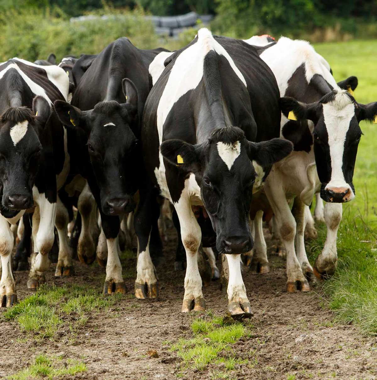
During a recent herd test reading of a dairy herd, my client Johns’ yearling heifers were coming out of the building, it was immediately obvious that one of the animals was severely lame. She was hardly able to leave her front leg on the ground.
A first examination in the crush showed some swelling around the fetlock and some unusual movement within the joint. As I handled the joint, it was difficult to determine exactly where the injury was located and if the joint itself was involved. In comparison to other broken bones I has seen over the years – this one just felt “strange”.
Further Investigation
As the client had planned to keep the heifer for breeding and milking, we decided to investigate further and “not just put a cast on”. So after finishing the reading, I continued to the clinic and he loaded the animal onto the trailer and followed.
After sedation we positioned the heifer on the ground and I took some x rays. The result was rather surprising. There was no break, but instead, the whole fetlock was dislocated.
The groves and tuberositae of the joint “sitting beside where they should be” (see x ray to the left). Having the grove of the one bone and the tuberosita of the neighbouring bone on top of each other, repositioning proved difficult.
Manual Repositioning of the Joint
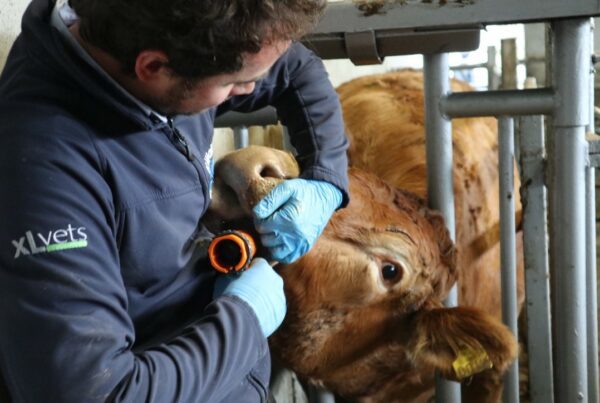
Three people were required to get the joint back in the right position. While Johns’ brother secured the heifer, John pulled on the foot with all his strength. This intense pressure was necessary to open the joint so we could move the tuberosita out of the grove. As you can imagine, without heavy sedation of the heifer, this method would have been impossible.
The heifer would have resisted the manipulation and because of the pain, the muscle would have been contracted instead of relaxing and without this relaxation any attempt would have been ineffective from the start.
While they held and pulled, I applied lateral pressure to the joint. Several attempts failed and while John was already wondering if we should give up, I gave it one more go. Putting all pressure possible on the joint, all of a sudden, I could feel the bones give under my hands and slip back into position. Immediately, we took a second x ray to confirm the result. (see x ray N° 2 to the left).
Aftercare
To stabilise the joint, and prevent sideways movement, I constructed a splint and secured it with a tight bandage of Vetwrap. Before the animal woke up again, I injected some pain-relief/anti-inflammatory drugs and supplied enough for the coming days.
I instructed John to restrict the heifer’s movement and aim to reduce stress from other animals of the group. The splint was removed after a week. At this point the swelling had gone down completely and lameness had significantly improved. Another supportive bandage was applied and was left on for an additional two weeks. After taking the bandage off and confirming a stable joint, the heifer was reunited with her friends!
While routinely x rays are not used in our clinic for large animals, we went through with it in this case. Only the x- ray revealed the true diagnosis, a complete dislocation of the fetlock joint.
As manual repositioning in a grown cow/bullock would have been impossible, for this young heifer it worked out just fine and she made an excellent recovery.